The American Roentgen Ray Society (ARRS) first traveled to San Diego, CA, for the 1996 ARRS Annual Meeting. Almost three decades ago, Kay H. Vydareny of Emory University Hospital in Atlanta, GA, was named this society’s first female president on Sunday, May 5. (Apropos, of the four newly elected 2025–2026 ARRS officers installed on Sunday, April 27, 2025 at the Marriott Marquis Marina, half are women.)
The latest convening of North America’s first radiological society delivered the same clinically relevant experience for which ARRS has long been heralded: radiologists of each practice type and every training level relishing world-class instruction from trusted experts spanning every subspecialty. Pioneers in asynchronous continuing education, ARRS continues to offer both inperson and virtual registrants the most flexible meeting experience in radiology. All attendees retain on-demand access to the complete program for an entire calendar year, learning and earning CME well into 2026.
And speaking of 2026, ARRS looks forward to delivering yet another singular experience in Pittsburgh, PA. This city of steel and bridges has reinvented itself as a hub of health care innovation, making David L. Lawrence Convention Center the perfect host for #ARRS26. Like the city itself, ARRS is constantly evolving— offering new education, fresh perspectives, and invaluable opportunities to connect with peers in meaningful ways. Join us to be part of a dynamic meeting where you’ll gain knowledge, build connections, and experience the energy of a city that’s shaping the future of patient care.

Opening Ceremony Honors Distinguished Educator, ARRS Scholars, and 2025 Gold Medalist
After ratifying an amendment to Article X of the bylaws to clarify that ARRS now publishes two radiology journals—AJR and R3—the ARRS membership officially installed Deborah A. Baumgarten, MD, MPH, of the Mayo Clinic in Jacksonville, FL, as the 125th president of ARRS. An internationally recognized leader in radiology and a steadfast advocate for education, Dr. Baumgarten assumes the presidency after years of distinguished service on the ARRS Executive Council.
Her appointment marks a new chapter in this society’s ongoing mission to advance medical imaging and patient care through expert education, cuttingedge research, and fruitful collaboration. Widely published and highly respected for her clinical expertise, instructional insights, and leadership within the imaging community at large, Dr. Baumgarten succeeds Angelisa M. Paladin, MD, who presented the ARRS presidential gavel to her successor.
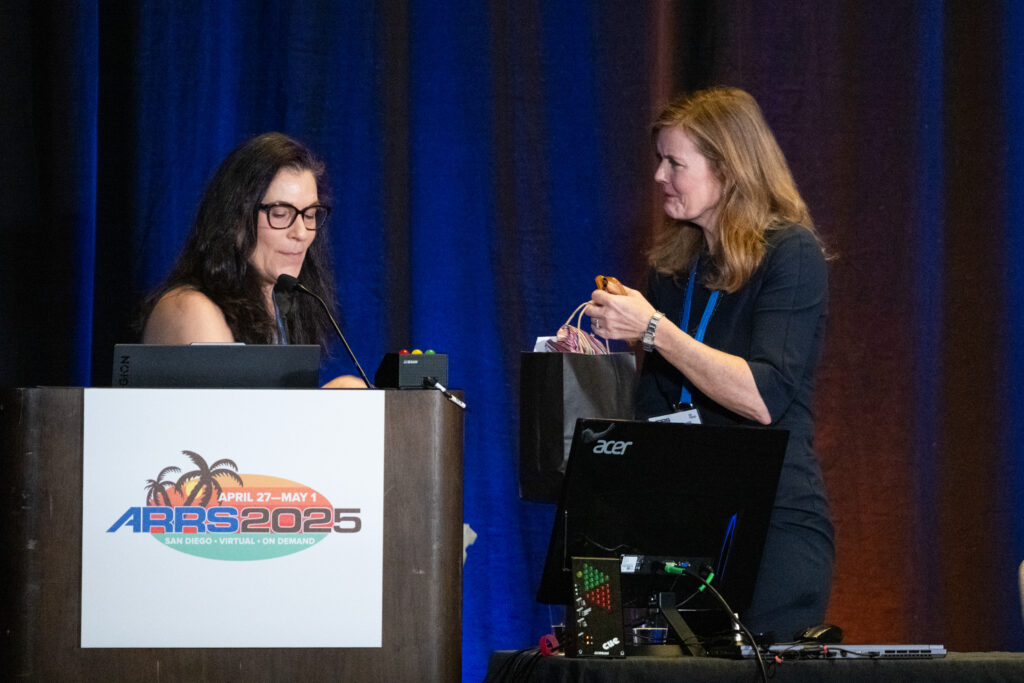
First, Tanya W. Moseley, MD, of the University of Mississippi Medical Center was honored as the 2025 ARRS Distinguished Educator. For more than 20 years, Dr. Moseley’s contributions to ARRS have transformed radiological education. Through her service on key educational committees and roles as casebased breast imaging chair and AJR SA-CME Consultant Editor, she has fundamentally shaped the organization’s educational direction. Her pioneering vision led to the creation of the first multi-vendor tomosynthesis certification course, demonstrating her exceptional ability to build collaborative educational programs. As architect and director of the ARRS Longitudinal Course Series, she continues to advance innovative approaches to radiology education.

This society was then doubly proud to recognize two recipients of 2025 ARRS Scholarships: Matthew Lee, MD, at the University of Wisconsin School of Medicine and Public Health and Yale School of Medicine’s Luca Pasquini, MD, PhD. Provided by The Roentgen Fund, ARRS Scholarships support early-career faculty members pursuing radiological research that promises to change how medical imaging is practiced. A two-year grant totaling $180,000, the ARRS Scholarship aims to advance emerging scholars, as well as prepare them for positions of leadership.

The biggest laurel of the morning, the ARRS Gold Medal, went to Ruth C. Carlos, MD, MS, FACR. Installed as president of ARRS during the 2019 Annual Meeting in Honolulu, HI, presently, Dr. Carlos is a professor at Columbia University Irving Medical Center and associate chair of research faculty development for the radiology department. And as fellow past ARRS president and longtime Michigan colleague N. Reed Dunnick introduced her: “Ruth Carlos isn’t just a superstar. I’d argue that she’s an entire galaxy!” Indeed, having served 11 years on our Executive Committee, as well as 11 ARRS committees prior, Dr. Carlos remains a fixture in our universe. There’s no one more deserving of the highest merit bestowed by this society, which has been honoring distinguished service to radiology for more than four decades.
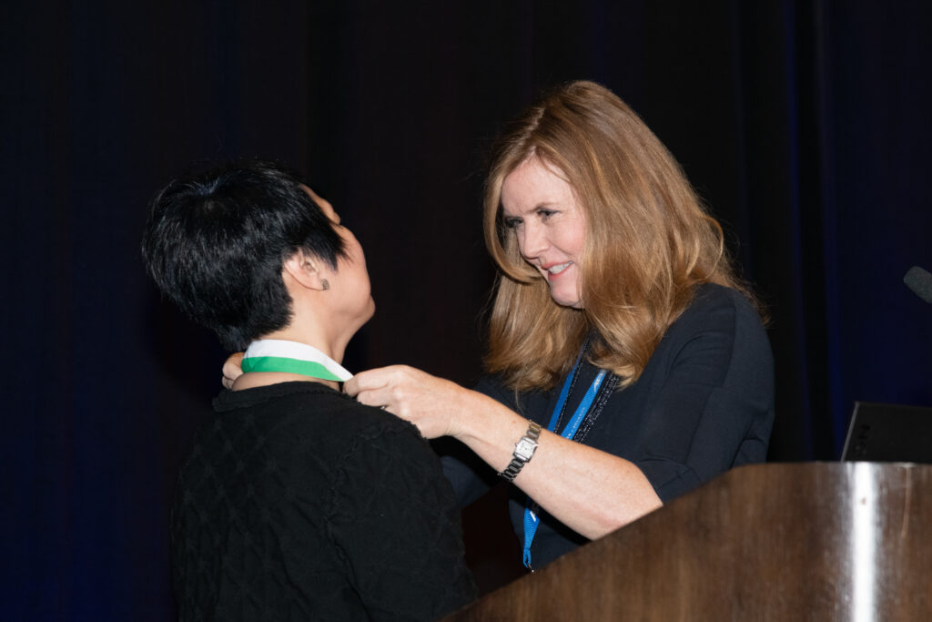
Service Over Self: Longtime ARRS ED Receives New Lifetime Award
The entirety of the ARRS was eager to give the first-ever ARRS Lifetime Service Award to its former executive director Susan B. Cappitelli, MBA, CAE. “For many of us who have been attending the ARRS Annual Meeting for many years,” Dr. Paladin remarked, “you’ve seen a huge growth in our programming and our services.” Well, that growth started with Susan way back in 1991, when ARRS recruited her to bring every aspect of producing the world’s longest published general radiology journal, AJR, in house. Needing a home for all the talented and dedicated professionals Susan herself was recruiting, six years later, she helped ARRS secure its own office building in Leesburg, VA. The society would go on to use every inch of space, of course, because during Susan’s historic tenure as executive director, ARRS membership increased more than 155%. Meanwhile, the society’s net assets ballooned from just $5 million to over $30 million. Having steered the membership and organization through everything from RVUs to COVID and beyond, “Susan always led with grace,” Dr. Paladin added, “and she will never, ever be forgotten.”
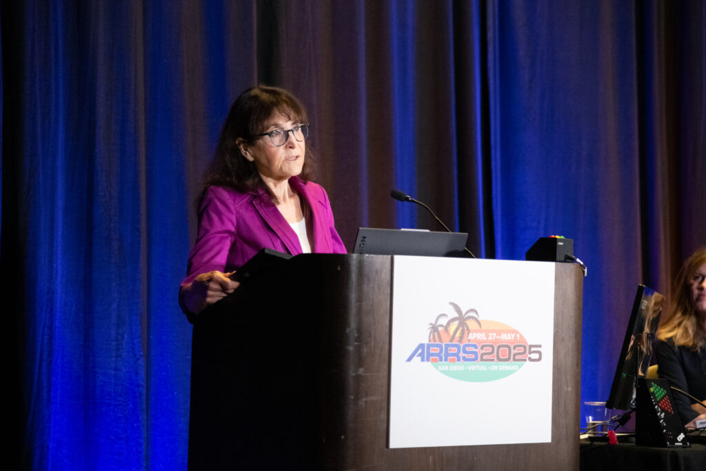
Welcome to 2025 Honorary ARRS Member, Salvador Amézquita Pérez
Dr. Salvador Amézquita Pérez, president of Sociedad Mexicana de Radiología e Imagen (Mexican Society of Radiology and Imaging), received his honorary ARRS membership on day one, and he and his colleagues were welcomed to the 2025 ARRS Annual Meeting in San Diego as part of the Global Exchange Featuring Mexico. The ARRS-SMRI Sunday Featured Course, “Advances in Cardiac Imaging,” focused on techniques and considerations for evaluating structural heart conditions and coronary artery anomalies. This session addressed non-atherosclerotic coronary artery narrowing, CT’s role in transcatheter mitral valve replacement and adult congenital heart disease, CT perfusion and FFR-CT for myocardial ischemia, as well as CT in TAVR before and after surgery.
The mission of the ARRS Global Partner Society Program is to build long-standing relationships with key leaders and organizations in the worldwide imaging community—increasing awareness of our society’s services in specific nations, while raising the stature of Global Partner Societies among ARRS members. Every year, the ARRS Annual Meeting Global Exchange incorporates one partner society into the educational and social fabric of our meeting. ARRS members then reciprocate at the partner society’s meeting that same year. Our 2026 Annual Meeting Global Exchange will welcome a delegation from the Royal Australian New Zealand College of Radiologists to Pittsburgh, PA.
AJR Luncheon Recognizes 2025 Figley and Rogers Journalism Fellows
During the American Journal of Roentgenology (AJR) Luncheon on Monday afternoon, Erin Alaia, MD, of NYU Langone Health in New York City was honored as the 2025 Melvin M. Figley Fellow in Radiology Journalism. Domen Plut, MD, PhD, from Slovenia’s University Medical Centre Ljubljana was recognized as the 2025 Lee F. Rogers International Fellow in Radiology Journalism.

Named for two distinguished Editors Emeriti of AJR, the Melvin Figley and Lee Rogers Fellowships offer practicing radiologists an unparalleled opportunity to learn the tenets of medical publishing via “the yellow journal”—the world’s longest continuously published radiology journal. Through hands-on experience with ARRS staff and AJR personnel—as well as personal apprenticeship with AJR’s 13th Editor of Chief, Andrew B. Rosenkrantz— Drs. Alaia and Plut will receive expert instruction in scientific writing and communication, manuscript preparation and editing, peer review processes, journalism ethics, and digital production and publication.
With Distinction: Award-Winning Scientific Research Presented in San Diego
The 2025 ARRS Annual Meeting hosted hundreds of electronic exhibits and abstracts presenting leading-edge research. Below are but a few highlights from our award-winning Scientific Program posters.
Mitigating Disparities in MASLD—Advancing Early Image Detection and Management

According to the Summa Cum Laude Award-Winning Online Poster presented during the 2025 ARRS Annual Meeting, patients with metabolic risk factors and an image diagnosis of steatosis— but without a recognized diagnosis of metabolic-associated steatotic liver disease (MASLD)—were more likely to experience significant complications of chronic liver disease, including cirrhosis and hepatocellular carcinoma (HCC), when compared to patients with a formal MASLD diagnosis. A public health crisis with significant morbidity and mortality implications, “MASLD affects approximately one-fourth of the global population, making it the most chronic liver disease worldwide,” noted lead investigator Emmanuel Mgboji of the University of Michigan Medical School. Using data from his institution’s EMR, Mgboji and fellow Michigan researcher Jessica Fried, MD, identified a cohort of 10,280 subjects with a formal diagnosis of MASLD and 5,103 subjects without a formal MASLD diagnosis but with MRI evidence of hepatic steatosis and metabolic risk factors. After demographic extraction and comparison, Mgboji and Fried monitored each cohort for progression of disease-related outcomes from 2018 to 2023, including HCC, cirrhosis, myocardial infarction (MI), chronic kidney disease (CKD), coronary artery disease (CAD), and type 2 diabetes mellitus (T2DM). Then, after comparing the incident rate and relative risk for each cohort, subjects were stratified by race (n = MASLD Dx; n = Image Dx cohort), including Caucasian Americans (CAs) (n = 8,548; n = 4,227), African Americans (AAs) (n = 580; n = 399), Asian American (ASAs) (n = 599; n = 229), and Hispanic Americans (HA) (n = 433; n = 169). “Comparing incidence rates between the MASLD Dx and Image Dx cohorts (no MASLD Dx cohort),” Mgboji said, “we found a significant relative risk of 2.185 (95% CI: 1.6822–2.8395, p < 0.0001) for a diagnosis of HCC in the Image Dx cohort. Additionally, the relative risk of developing cirrhosis in the Image Dx cohort was 1.459 (95% CI: 1.2934–1.6461, p < 0.0001).” Mgboji and Fried also assessed the risk of being diagnosed with MI, CKD, CAD, and T2DM in the Image Dx cohort, with a relative risk of 1.236 (p = 0.0496), 1.240 (p = 0.0002), 1.346 (p < 0.0001), and 1.259 (p < 0.0001), respectively. “Further significant differences were observed when patients were stratified by racial groups,” added Mgboji. For developing HCC, the relative risk was 2.465 (p < 0.0001) in CAs, 3.488 (p = 0.0180) in AAs, 1.962 (p = 0.375) in ASAs, and 4.270 (p = 0.0451) in HAs. For cirrhosis, relative risk values were 1.590 (p < 0.0001) for CAs, 1.817 (p = 0.0248) for AAs, 1.933 (p = 0.0336) for ASAs, and 2.795 (p = 0.0003) for HAs.
BI-RADS 3 “Report Card” Decreases the Rate of Usage

In the Magna Cum Laude Award- Winning Online Poster at this year’s Annual Meeting, anonymous, peer comparison BI-RADS 3 “report cards” proved to be an effective method of rate reduction, particularly at community hospitals where preintervention rates were higher than at academic sites. Reserved for “probably benign” breast imaging abnormalities that have a low (< 2%) risk of being malignant, in practice, the actual use of ACR’s BI-RADS category 3 assessment varies among radiologists— often overutilized to equivocate a finding. Giving radiologists a recommended target rate of less than 12% as a benchmark, head presenter Bonmyong “Bora” Lee, MD, and her team of researchers from UPenn’s Perelman School of Medicine sent quarterly BI-RADS 3 report cards to each breast imaging radiologist via automated emails. This report card included personal BIRADS category 3 rates for each modality, as well as cumulative rates for the radiologist’s covering site and hospital. Each radiologist was blinded to others’ individual rates, participation was voluntary, and Lee et al. offered neither rewards nor punitive measures for performance. Noting that radiologists were not monitored for review compliance either, “after 4 cycles, we reviewed the data to determine if there were changes in the rate of BI-RADS 3 assessment among radiologists and across the institution using paired t-tests,” Lee said. Over Lee et al.’s 17-month assessment period, 38 radiologists issued BI-RADS 3 in 4,289 total patients: 1,171 diagnostic mammograms, 1,281 screening mammograms, 658 MRI, and 1,179 ultrasound examinations. After Lee and colleagues’ intervention, the average BI-RADS 3 rate decreased (all sites: p < 0.01; community sites: p < 0.01; academic sites: p = 0.07). Radiologists with preintervention BI-RADS 3 rates that were greater than the group median had larger reductions in BI-RADS 3 rates post-intervention (p < 0.05).
Significance of Nonspecific 18F-DCFPyL Rib Uptake on PET/CT in Prostate Cancer Patients

The Cum Laude Award-Winning Online Poster concluded that evaluating increased rib uptake on 18F-DCFPyL PET/CT may be challenging, even resulting in false-positive findings. “In patients without osseous metastasis, the uptake is often low (less than the mean liver SUV), stable, and likely represents benign etiologies, such as fibrous dysplasia, fibrous cortical defect, traumatic fractures, and hemangiomas. Thus, further evaluation is usually not required,” said presenter Aisha Alam, DO, from the Icahn School of Medicine at Mount Sinai in New York, NY. Dr. Alam and her all-Icahn School team performed a retrospective review of patients with prostate cancer who underwent PET/CT scans with radiotracer fluorine-18 2-(3-{1-carboxy-5-[(6-18F]fluoro-pyridine- 3-carbonyl)-amino]-pentyl}-ureido)-pentanedioic acid (18F-DCFPyL) at a single academic center from June 2021 to April 2024. Alam and colleagues’ EHR search identified patients with increased radiotracer rib uptake. The team excluded patients with imaged evidence of osseous metastasis at other sites or with CT suggestive of alternate diagnoses, then analyzed Gleason scores, most recent serum PSA levels, maximum standardized uptake values (SUVmax) in the rib foci, and mean liver SUV. Finally, for confirmation of benignity, follow-up evaluation included stability on 18F-DCFPyL PET/CT and/or imaging modalities such as CT, bone scan, or MRI. With 204 total 18F-DCFPyL PET/CT scans showing solitary or multiple foci of increased rib uptake without CT correlation and imaging evidence of osseous metastatic disease, the mean age was 68 years old, mean Gleason score was 7, mean PSA was 7.9 ng/mL (range: undetectable–50.2 ng/mL), mean SUVmax for rib uptake was 3.8 (range: 1.4–9.8), and mean liver SUV was 5.7 (range: 2.7–10.9). “Of the 204 scans,” Alam noted, “31 studies belonged to 13 patients who underwent follow-up 18F-DCFPyL PET/CT for restaging.” For patients with redemonstrated rib uptake, mean SUVmax was 3.5 and mean SUVmax percent change on subsequent scans was 15.1% (range: 0–46.4%). And for patients with multiple PET/CT scans, the SUVmax for each foci of rib uptake was less than the mean liver SUV (mean: 6.1, range: 4.0–8.8). Of the 23 single-scan PET/CT patients who had a rib uptake greater than the liver mean, 14 of those had a PSA less than 10 ng/mL. “Larger studies evaluating similar findings with histologic correlation and follow-up imaging are needed to improve diagnostic certainty,” added Alam et al.
Analysis of Breast Radiation Therapy and Breast Arterial Calcifications on Screening Mammography

Findings from a Certificate of Merit Online Poster presented during ARRS 2025 suggest that breast radiation therapy exposure does not impact the prevalence of mammographic breast arterial calcification— therefore, not impacting its utility as an imaging biomarker of cardiovascular disease risk. “Our study is the first retrospective analysis of the association between breast cancer radiation therapy exposure and the presence of breast arterial calcification on screening mammography,” noted presenter Jessica Rubino, MD, from Dartmouth Hitchcock Medical Center in Lebanon, NH. Rubino et al. performed an electronic health database query to identify women ages 40–75 years who had a screening mammogram between January 1, 2011 and December 31, 2012. After a chart review to extract data regarding breast cancer radiation therapy history, two breast imaging radiologists then reviewed mammograms for the presence of breast arterial calcification. The researchers used multivariate logistic regression to examine the association between breast radiation therapy exposure and breast arterial calcification, adjusting for age, BMI, smoking status, hypertension, type 2 diabetes, as well as use of statin and antihypertensive medication. Of the 1,155 women included in this analysis, 222 (19.2%) had mammographic evidence of breast arterial calcification, 122 (10.6%) had a history of radiation therapy exposure, and 39 (32%) women with radiation therapy exposure had breast arterial calcification on the index mammogram obtained at least 2 years after completing radiation therapy. Compared to women without radiotherapy, women with a history of breast radiation therapy exposure had higher odds of breast arterial calcification (OR: 2.18, 95% CI: 1.43–3.28; p = 0.0008). After multivariable adjustment, however, this association became nonsignificant, with the maximally adjusted model demonstrating an OR of 1.52 (0.95–2.40; p = 0.07).